White nail
Mees line, Lindsay nails , Muehrcke lines (see below), and punctate leukonychia may be associated with:
Muehrcke’s Lines of the Fingernails
Muehrcke’s lines appear as double white lines that run across the fingernails horizontally.
The condition is named after Robert Muehrcke, MD. He first described the condition in the British Medical Journal in 1956.
Symptoms of Muerhcke’s Lines
Muehrcke’s lines usually affect several nails at a time. There are usually no lines on the thumbnails.
Some characteristics of Muehrcke’s lines are:
- White bands go across the entire nail from side to side.
- Lines are usually most clearly seen on the second, third, and fourth fingers.
- The nail bed looks healthy in between the lines.
- The lines do not move as the nail grows.
- The lines do not cause dents in the nail.
- When you press down on the fingernail, the lines temporarily disappear.
Causes of Muehrcke’s Lines
The exact cause of Muehrcke’s lines is not clearly understood. The lines are not caused by injury to the cuticle or nail area.
The lines have been linked to low levels of a protein called albumin. Albumin is found in the blood. It is made in the liver.
Albumin plays an important role in many body functions. It keeps fluid from leaking out of your blood vessels. It also helps move hormones, vitamins, and medicines through your body.
Although low albumin level is most commonly linked to liver disease, many different systemic (body-wide) diseases can cause low albumin levels. Muehrcke’s lines have been seen in people with:
- Cancer after chemotherapy
- Kidney disease, including nephrotic syndrome and glomerulonephritis
- Liver disease, including cirrhosis
- An unbalanced diet that leads to an extreme lack of nutrients in the body (severe malnutrition)
One possible theory for the appearance of Muehrcke’s lines is that these diseases lead to swelling in the nail bed. The swelling puts pressure on the blood vessels that run underneath the nail, causing color changes.
The lines have also been seen in older people receiving chemotherapy who have normal albumin levels. However, Muehrcke’s lines most often occur in those with too little albumin.
Treatment of Muehrcke’s Lines
If your albumin level is too low, you may be given albumin through a vein (by IV, or intravenously).
The lines tend to go away when your albumin level returns to normal, or near normal. However, a normal range may vary, depending on the lab that tested your blood. Always talk to your doctor about your test results.
Other treatment depends on the disease or disorder that caused the fingernail changes.
Show Sources
American Family Physician Web site: “Nail Abnormalities: Clues to Systemic Disease.”
The American Journal of Medicine Web site: “Muehrcke’s Lines.”
Lab Tests Online web site. “Albumin: The Test.”
Scher, R.K. Scher & Daniel: Nails: Diagnosis, Therapy, Surgery, 3rd ed., Saunders Elsevier, 2005.
James, W.D, Berger, T.G., Elston, D.M., eds. Andrews’ Diseases of the Skin: Clinical Dermatology, 11th ed., Saunders Elsevier, 2011.
Merck Manual for Health Care Professionals Web site: “Acrocyanosis.”
Muehrcke, R. British Medical Journal, 1956.
The American Journal of Medicine, November 2010.
Morrison-Bryant, M. New England Journal of Medicine, 2007.
White nail
Author(s): Linda Mardiros, University of Ottawa, Canada; Hana Numan, Senior Medical Writer, DermNet (2022)
Previous contributors: Hon A/Prof Amanda Oakley, Dermatologist (2016)
Reviewing dermatologist: Dr Ian Coulson
Edited by the DermNet content department
Table of contents
arrow-right-small
What are white nails ?
A white nail , also known as leukonychia , is the partial or full discolouration of the nail plate on one or more fingernails or toenails.
White nails are the most common nail dyschromia. The nail will lose its general pink undertone and appear white.
White nail

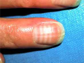
Parallel bands of leukonychia due to cyclical chemotherapy
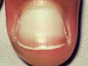
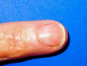
‘Half and half nail’: leukonychia is affecting the proximal half of the nail
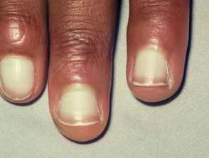
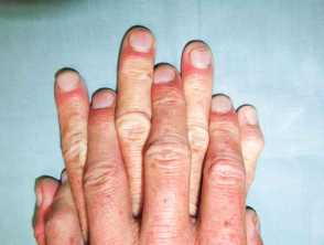
Leukonychia due to hypoalbuminaemia
How is a white nail classified?
Leukonychia can be classified by underlying pathology , its distribution , or how it develops.
Classification according to pathology
Leukonychia can be subdivided into true and apparent discolouration.
- True leukonychia: discolouration due to abnormal nail plate keratinisation . The white nail will not be hidden by pressure application of the nail plate to the bed.
- Apparent leukonychia: secondary to disease of the nail bed. This appearance disappears with pressure application on the nail.
Classification according to distribution
Leukonychia can be partial or total.
- Total leukonychia: whitening of the entire nail plate.
- Partial leukonychia: 3 subtypes are described.
- Punctate
- Longitudinal
- Striate (see below).
Classification by development timeline
White nails can be acquired or congenital .
- Congenital: familial leukonychia is more commonly inherited recessively, although dominant patterns are possible. This is due to a mutation in the phospholipase C delta-1 gene in which all nails appear milky and porcelain white.
- Acquired: secondary to systemic disease. Important to note that congenital leukonychia may also be secondary to systemic disease (see below).
Who gets a white nail?
White nails can affect anyone of any gender , age or ethnicity. Its presence may warrant a work-up for systemic disease.
What causes a white nail?
Trauma
- True leukonychia: partial or whole nail plate damage caused by injury to the nail plate or matrix. Keratin disruption with trapped air within the nail plate, resulting in reflection and lack of transparency.
- Punctate leukonychia: occurs after nail biting, manicuring, knocks and bangs, and tight footwear use.
- Striate leukonychia: also known as Mees lines or transverse leukonychia, may follow damage to the nail matrix ; furrows and ridges may also appear.
- Total leukonychia: can follow a more serious injury, often with detachment of the nail plate from the nail bed, and alteration to the nail contour.
Poisoning and drugs
Mees line, Lindsay nails , Muehrcke lines (see below), and punctate leukonychia may be associated with:
- Heavy metal poisoning (eg lead, arsenic)
- Chemotherapy
- Sulphonamides.
There are three distinct types of apparent leukonychia that may be associated with the systemic disease:
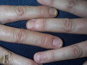
- Muehrcke lines: A pair of observable, non- palpable , horizontal (transverse) white lines across the nail due to variable blood flow
- Lindsay nails (half-and-half nails): Proximally white or pink-coloured nail with a distal darkening
- Terry nails : Whitening of the majority of the nail with a thin 0.5-3.0 mm distal darkening.
Systemic illness
Terry nails have been associated with:
- Liver cirrhosis
- Chronic kidney disease
- Heart failure
- Hypoalbuminemia (due to protein malabsorption, eg, in colitis )
- Protein-losing enteropathy
- Diabetes
- Iron deficiency ( anaemia )
- Zinc deficiency
- Hyperthyroidism .
Lindsay nails have been associated with:
- Chronic kidney disease
- Psoriasis.
Muehrcke lines have been associated with:
What are the clinical features of a white nail?
True leukonychia with partial distribution:
- Punctate leukonychia:
- Most common form of true leukonychia
- Described as small white spots on the nails
- Tends to be the result of trauma and isolated to a few nails.
- Small white longitudinal bands
- Presenting in individuals with Darier disease or Hailey-Hailey disease.
- One or more white horizontal bands across the entire nail in parallel with the lunula .
Patients with multiple true leukonychia warrant a thorough history, physical examination, and medication review to exclude a toxic or systemic etiology. This is also true of leukonychia extending the full width of the nail plate.
How do clinical features vary in differing types of skin?
They appear similar in different skin types.
What are the complications of a white nail?
White nails are a cosmetic nuisance but may be a marker of an underlying systemic disease, but per se do not have any physical complications.
How is a white nail diagnosed?
A thorough history and physical examination may be sufficient for diagnosis. Although, when the cause is unclear, the following tests may be helpful:
- Nail clippings to exclude fungal infection
- Nail biopsy
- Blood tests to evaluate systemic disease, particularly renal and liver function tests.
What is the differential diagnosis for a white nail?
- Onychomycosis (also known as pseudoleukonychia) — the disease of the nail plate is due to external factors
- Onycholysis
- Nail psoriasis
- Trachyonychia (twenty-nail dystrophy )
- Vitiligo of the nail.
Onychomycosis
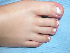
White discolouration of the distal nail plate in superficial white onychomycosis
What is the treatment for a white nail?
Treatment ultimately depends on the presence of any underlying cause.
There is no treatment for trauma-related leukonychia. Punctate lesions will disappear as the nail follows its natural growth pattern (around 6 to 9 months for a fingernail).
How do you prevent a white nail?
Avoidance of trauma-induced leukonychia will help prevent development of white nails.
- Avoiding contact with irritating substances, and wearing appropriate protective equipment if contact is required.
- Avoiding excessive use of nail polish or excessive mechanical force with false nail application/removal.
- Minimising picking and biting nails.
- Wearing appropriate shoes to prevent excessive pressure on toes.
- Applying moisturisers to hydrate nails.
What is the outcome for a white nail?
Leukonychia due to minor trauma or medication may completely resolve over a few months. In other cases, the white nail plate may remain permanently or demonstrate recurrence .


